Mayson H Alkhatib1 ![]() ,
Huda M Alkreathy2,
Khadijah S Balamash1,
Faiza Abdu3
,
Huda M Alkreathy2,
Khadijah S Balamash1,
Faiza Abdu3
For correspondence:- Mayson Alkhatib Email: mhalkhatib@kau.edu.sa Tel:+966599240526
Received: 28 December 2015 Accepted: 17 April 2016 Published: 27 May 2016
Citation: Alkhatib MH, Alkreathy HM, Balamash KS, Abdu F. Antitumor activity of doxorubicine-loaded nanoemulsion against Ehrlich ascites carcinoma-bearing mice. Trop J Pharm Res 2016; 15(5):937-943 doi: 10.4314/tjpr.v15i5.6
© 2016 The authors.
This is an Open Access article that uses a funding model which does not charge readers or their institutions for access and distributed under the terms of the Creative Commons Attribution License (http://creativecommons.org/licenses/by/4.0) and the Budapest Open Access Initiative (http://www.budapestopenaccessinitiative.org/read), which permit unrestricted use, distribution, and reproduction in any medium, provided the original work is properly credited..
Purpose: To evaluate the antitumor activity of doxorubicine (DOX)-loaded nanoemulsion (NE) on Ehrlich ascites carcinoma (EAC)-bearing Swiss albino mice.
Methods: The mice were divided into five groups (n = 20) according to the administered drug. Groups I - V were labeled as negative control (normal), positive control of the untreated EAC bearing mice (EAC control), blank nanoemulsion (BI-NE), DOX-loaded-NE (DOX/LNE) and free DOX (DOX-Sol), respectively. Cardiotoxicity was assessed by measuring changes in body and organ weight, analyzing serum enzymes and lipids, and examining histological changes in heart tissues by light microscopy. In addition, mean survival time (MST), increase in life span (ILS) and survival (S) of the mice were determined.
Results: DOX/LNE group reduced levels of serum enzymes and lowered damage to heart tissues relative to DOX-Sol group. The MST of the DOX/LNE group (80 ± 0.0 days) was significantly greater than that for DOX-Sol group (34.6 ± 8.9 days), while ILS of DOX/LNE (265.30 days) was higher than that of DOX-Sol (57.99 days) by 4.6-fold.
Conclusion: Administration of DOX/LNE to EAC-bearing mice improves the efficacy of DOX and reduce its side effects on the heart.
Introduction
Cancer is a potential fatal disease, characterized by uncontrolled growth and spread of abnormal cells [1]. In spite of the spread of many anticancer drugs, there are still challenges in employing them in cancer therapy because of their poor bioavailability, water insolubility, high toxicity and nonspecific targeting which result in damaging the healthy cells. Doxorubicin (DOX) is one of the most effective anticancer drugs, with a wide scope of activity in human cancers, including acute lymphoblastic leukemia, breast carcinoma, ovarian carcinoma, and hepatocellular carcinoma [2]. However, its clinical application is limited by its harmful side effects, the most significant of which is its cardiotoxicity that can lead to cardiomyopathy and congestive heart failure [3].
A variety of approaches have been reported to minimize the effective chemotherapeutic dose and thereby its side effects. It is possible to devise nanocarriers to deliver anticancer drugs which would allow efficient passage and high bioavailability of the anticancer drugs. Nanoemulsions (NE) are one of the promising nanocarriers for many drugs [4]. They are non-equilibrium, heterogeneous systems consisting of two immiscible liquids and mixed surfactants and cosurfactants which facilitate the dispersion of one liquid in the other as droplets with diameters ranging from 1 to 100 nm [5]. Anticancer drugs loaded in NE systems have greater activity against cancer cells in comparison to other emulsification systems. This is due to the decreased particle size and zeta potential, production of a stable water dispersion, reduced polydispersity index, and greater stability of drug with the NE [6].
In the current study, the anticancer activity of the DOX/LNE on the EAC bearing mice was elaborated.
Methods
Materials
DOX hydrochloride was obtained from Pharmacopeia, California, US. Soya phosphatidylcholine (SPC), sodium oleate (SO), tris (hydroxymethyl) amino methane, polyoxyethyleneglycerol trihydroxystearate 40 (Eumulgin HRE 40, EU) and hydrochloric acid (HCl) were purchased from Leo Chem., S. Puran, Bangalore, India. Cholesterol (CHO) was purchased from Jechno pharmacies BanadurGarh, Haryana, India. 1-Octanol was obtained from AflaAesar GmbH & Co KG Karlsruhe, Germany.
Subjects
Healthy female Swiss albino mice weighing 25 – 30 g were used for the experiment. The animals were housed in large polypropylene cages with utmost of five animals per cage and maintained under standard laboratory conditions (temperature 25 ± 2 °C with dark/light cycle 12/12 h) and provided with standardized pelleted feed and clean drinking water ad libitum. The animals were maintained in accordance with King Abdulaziz University's policy and international guidelines on the care and use of laboratory animals [7]. Ethical approval was obtained from Research Ethics Committee of Faculty of Medicine at King Abdulaziz University (approval ref. no. 189/247/1433). Ehrlich ascites carcinoma (EAC) cells were obtained from American Type Tissue Culture Collection (Manassas, VA, USA).
Preparation of nanoemulsion formulations
The nanoemulsion (NE) formulation was prepared as described elsewhere [8]. In brief, a 1.25 % (wt/wt) of the solid surfactants, EU/SPC/SO, were mixed at fixed ratio of 3.5:3.0:3.5. Then, a 0.125 % (wt/wt) of the oil phase of CHO was added slowly to the surfactant mixture until a semisolid phase was formed. After that, a 1 % (wt/wt) of 1-octanol was added dropwise. Finally, the mixture was diluted with the aqueous phase of 97.63 % (wt/wt) of warm Tris–HCl buffer (pH 7.22). The mixture was kept in the water bath at 75 °C for three hours to be clear and transparent and the resulted NE formula was stored at 25 °C. The NE formulas were BI-NE, DOX/LNE, prepared by solubilizing 2.0 mg of DOX/ kg of mice in 0.2 ml of NE. The DOX-Solution (DOX-Sol) was prepared by dissolving 2.0 mg of DOX in 0.2 ml of distilled water.
In vivo antitumor activity
Transplantation of tumor and experimental protocol
The in vivo evaluation of the antitumor activity of different drug formulations was performed by using the regimen of Alkreathy et al [9]. The EAC cells were maintained in the ascitic form by sequential passages in the mice, by means of intraperitoneal (i.p.) transplantation of 2.5 × 10⁶ cells/mouse every 10 days. Ascitic fluid was drawn out from EAC bearing mice at the 8th day following transplantation. Before injection, the viable EAC cells, collected from the peritoneal cavity, were counted by trypan blue exclusion test.
The mice were divided into five groups (n = 20) according to the administered drug, at which Group I-V were labeled as negative control (normal), positive control of the untreated EAC bearing mice (EAC control), BI-NE, DOX/LNE and DOX-Sol, respectively. The body weight of all mice was taken before injection with EAC cell. All of the mice in each group were injected i.p. with 2.5 × 10⁶ EAC cells/mouse at day zero, except group I which was injected with saline solution. After 24 h, animals in Groups III-V were injected with 3 doses of the desired drug, every other day.
On the 15th day, ten mice from each group were fasted for 24 h and the body weight of each mouse was taken on a digital scale. Then, the blood was collected by a retro orbital plexus method for the estimation of serum biochemical assays. Finally, these animals were sacrificed for histology investigation. The rest of the animals in each group were kept for the survival study.
Relative organ weight
Immediately following sacrifice, the hearts of all of the mice were removed, washed with buffer and dried on blotting paper. Then all hearts were weighed on a digital scale. To compute the organ weight-to-body weight ratio, the post-sacrifice organs (heart) weight (B) from the same animal was divided by the pre-sacrifice body weight for each animal (A).
Determination of biochemical parameters
For biochemical assays, the blood samples were allowed to clot and the serum was separated by centrifugation at 5000 RPM for 10 min. Serum was utilized for the estimation of cardiac enzymes (creatine phosphokinase (CK) and lactate dehydrogenase (LDH)) and the serum lipid profile (cholesterol (CHO), high density lipoprotein (HDL) and triglyceride (TG)). The biochemical assays were performed according to the standard kit methods using fully Automated COBAS® 8000 modular analyzer series in the King Fahd Armed Forces Hospital–Jeddah, Saudi Arabia.
Histological studies
The collected heart tissues were fixed in 10 % neutral buffered formalin (4 % formaldehyde in phosphate buffered saline). Thereafter, the tissues were incrementally dehydrated by increasing concentrations of alcohol and then stained with hematoxylin and eosin (H & E), followed by microscopic examination.
Survival study
The observation period of the survival study began in the first weeks of age until the study ended at the twelfth week of age. The MST, % ILS and % S of each group containing ten mice were monitored by recording the daily mortality for 80 days. ILS were calculated using Eq 1 [10].
ILS (%) = {(St/Sc) – 1}100 …………………. (1)
where St and Sc are the mean survival time of treated and control groups, respectively.
Statistical analysis
All values were expressed as mean ± standard deviation (SD; n = 10). Statistical analysis was performed with one-way analysis of variance (ANOVA) test and independent sample t-test using MegaStat Excel (version 10.3, Butler University). The significant differences between the tested samples were classified according to the p-value. If p < 0.001, the difference was considered very highly significant (***); p < 0.01 was considered highly significant (**); the difference was considered significant at p < 0.05 (*).
Results
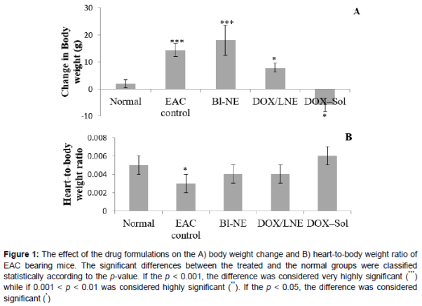
Body weight change and relative organ weight
As displayed in A, there was a very high significant increase in the body weight changes of EAC control and BI-NE groups compared with the normal group (P-value < 0.001) whereas the enhancement in the body weight change was reduced in the DOX/LNE group (p < 0.05). On the other hand, there was a significant decrease in the body weight change in the DOX–Sol group (p < 0.05). Studying the effect of the drug formulations on the heart weight relative to the body weight revealed that the mice treated with DOX–Sol, DOX/LNE and BI-NE showed no significant difference from the normal group in heart-to-body weight ratio, whereas EAC control group showed a significant decrease in the heart-to-body weight ratio (B).
Cardiac enzymes and lipid profile
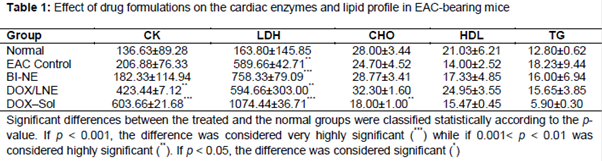
The effect of the drug formulations on the heart enzymes and lipid profile is exhibited in . The CK levels in the EAC control and BI-NE groups were comparable with the normal group. In spite of the significant increase in the CK levels in both of DOX–Sol and DOX/LNE relative to the normal group, the CK level of DOX/LNE group was significantly less than DOX–Sol group. Nevertheless, the CK levels in all of the groups were within the standard range of the enzyme in the mice (50-680 IU/l). Furthermore, all treated groups showed a significant increase in the LDH levels when compared to the normal group. However, the increase in the LDH levels in all of the groups were within the standard range of the albino mice, which is (85-750 IU/l), except DOX–Sol group which had elevated levels of LDH.
Regarding the lipid profile, there were no significant changes in CHO level between the normal group and the other treated groups except DOX–Sol group which had reduced amount of CHO. On the other hand, all treated groups did not show any considerable difference in HDL and TG levels when compared to the normal group.
Cardiac histopathology
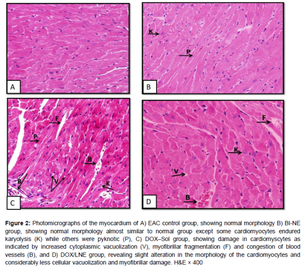
The histopathological examination of the heart tissues of mice in the EAC control group showed normal architectures as no inflammatory cell infiltration was seen (A). The tissue of the mice treated with BI-NE showed normal myocardium morphology almost similar to the EAC control group except some nuclei have endured karyolysis, which is known as the complete dissolution of the chromatin of a dying cell due to enzymatic degradation by endonucleases, while others were pyknotic, which is the irreversible condensation of chromatin in the nucleus of a cell undergoing necrosis or apoptosis followed by fragmentation of the nucleus (B). Mice treated with DOX-Sol showed damages in the heart tissues, including the loss of myofibrils (myofibrillar fragmentation), congestion of blood vessels, a stimulation of cytoplasmic vacuolization and some nuclei enduring karyolysis and pyknosis (C). DOX-LNE group had a significant reduction in the severity of myocardial degeneration that was observed as mild disruption of cardiac muscle fibers and reduced cytoplasmic vacuolization (D).
Survival
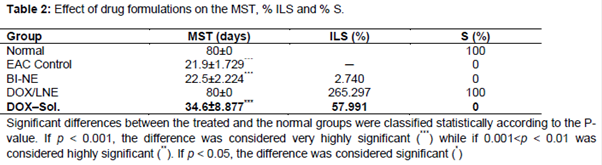
The effect of the drug formulations on the MST of EAC-bearing mice is illustrated in . Interestingly, the MST and % S of the DOX/LNE group was equivalent to the MST and % S of the normal group which was very highly significantly greater than the MST and % S of the EAC control, BI-NE and DOX-Sol groups. The % ILS of DOX/LNE was longer than the % ILS of DOX-Sol by 4.6 folds.
Discussion
The present study was carried out to evaluate the antitumor effect of the DOX-loaded-NE in one of the most devastating experimental tumor models, the EAC bearing mice. Because of their biocompatibility, NEs have been widely used in the medical field for delivery and controlled release of the hydrophobic anticancer drugs because they present a very large surface area and good long-term stability. As expected in the EAC control group, a significant increase in body weight was observed [10]. However, animals treated with DOX–Sol showed a further significant decrease in the body weight indicating more reduction in the i.p. tumor burden. This result was similar to several studies which demonstrated that the use of DOX leads to a significant drop in the body weight of rats [11,12]. In 2013, Lv et al [13] proved that DOX loaded in nanoparticles exhibited higher antitumor efficacy on A549 lung-tumor-bearing mice compared with free DOX at the same dose. Moreover, from our experimental groups, we found that DOX/LNE group had a significant decrease in the body weight compared to the EAC control group.
The reliable criterion for judging the value of any anticancer drug is the prolongation of life span of the tumor bearing animal [14]. The MST, % ILS and % S of EAC bearing mice treated with DOX/LNE were significantly longer than EAC bearing mice treated with DOX–Sol. This study is in agreement with Xiong et al [15] study, which showed that the life span of animals bearing MDA-435/LCC6WT (human breast cancer cell line) and treated with polymeric nanocarriers of DOX, was significantly longer than animals treated with free DOX. Additionally, Hao et al [16] demonstrated that nanoparticles can decrease the toxicity of DOX that result in a significant increase of the average lifetime in comparison with the free DOX.
In the present work, heart weight-to-body weight ratios were assessed in order to evaluate the toxicity of the drug formulas on the whole organ. Mice treated with drug formulations that contain 2 mg/ kg DOX did not show any significant change in heart weight-to-body weight ratio compared to the normal group. However, when rats were given three i.p injections of 5 mg/ kg DOX, a significant less mean ratio of heart weight to body weight against the control animals was reported [17].
DOX is a very potent antitumor antibiotic. Its use is severely limited for its cardiotoxicity, which have been documented in a variety of animal models [12]. During malignancy, the tumor marker enzymatic changes reflect overall changes in metabolism and they are increasingly appreciated as critical determinants of tumor cell behavior with sensitivity and specificity [18].
The results of the present study indicate that mice treated with DOX-Sol had a significant increase in LDH and CK levels, important markers of cardiac injury, especially during clinical follow-up of DOX therapy. In agreement with our findings, Yagmurca et al [19] demonstrated similar elevations in cardiac enzymes activities in rats following challenge with a single cumulative dose of DOX. Generation of free radicals by DOX caused extensive damage in the myocardium, which result in increased membrane permeability and hence leads to leakage of LDH and CK enzymes. On the other hand, administering DOX/LNE into the EAC bearing mice reduced the enhancement in the cardiac enzymes relative to the DOX-Sol. Park et al [20] proved that DOX-loaded-nanoparticle decreased the observed cardiac toxicity by decreasing CK level compared to the DOX treated group.
In the present study, it has been found that mice treated with Dox-Sol had significantly decreased levels in CHO compared to normal group. The levels of TG and HDL in mice were not affected by the different treated drug formulas. In contrast, rat treated with DOX at a total cumulative dose of 15 mg/kg through i.p showed that DOX interferes with metabolism and biosynthesis of lipids led to significantly increased levels of TG, CHO and decreased level of HDL [21].
According to the light microscopy images in the present study, heart sections of mice treated with DOX-Sol showed massive pathological changes compared to the normal architecture observed in the EAC control group. As observed in the heart tissue, the damages induced by DOX-Sol include the loss of myofibrils, congestion of blood vessels, stimulation in the cytoplasmic vacuolization and some nuclei enduring karyolysis and pyknosis. The histopathological changes observed in the DOX-Sol group were similar to those previously reported [22-24]. As noted from the histological reports, the heart myocytes degeneration could have resulted in the leakage of enzymes into the blood stream. Unlike DOX-Sol group, the heart tissue of DOX/LNE group was similar to the EAC control group sections in showing a normal tissue architecture. This result indicated that loading DOX in NE formulation reduced the damages and toxicity of DOX on the heart tissues.
Conclusion
The main goal of the current study was to assess the anticancer activity of DOX-loaded NE in vivo. Loading DOX into NE considerably improves its efficacy while reducing its cardiotoxicity. These findings suggest that NE is a promising nanocarrier for DOX. However, studies using other animals are required to confirm these findings and evaluate the possible toxicological effects of this new formulation in human patients.
Declarations
Acknowledgement
References
Archives


News Updates